Highlighted Antibody - PDL1 (Programmed cell death ligand 1) Clone BSR90
|
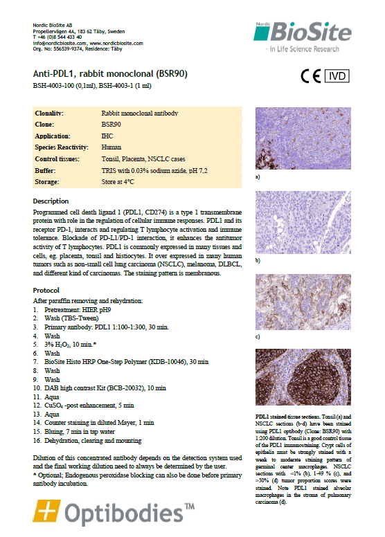
PDL1 is commonly expressed in many tissues and cells, eg. placenta, tonsil and histiocytes. It over expressed in many human tumors such as non-small cell lung carcinoma (NSCLC), melanoma, DLBCL, and different kind of carcinomas. The staining pattern is membranous.
This antibody provides truly optimal staining and as with all our antibodies we provide a 100% guarantee, so trying it really is risk free.
Suggested Protocol after paraffin removal and rehydration:
1. Pretreatment: HIER pH9 2. Wash (TBS-Tween) 3. Primary antibody: PDL1 1:100 – 1:300, 30 min. 4. Wash 5. 3% H2O2, 10 min 6. Wash 7. HRP One-Step Polymer, 30 min 8. Wash 9. Wash 10. DAB high contrast Kit, 10 min 11. Aqua 12. CuSO4 -post enhancement, 5 min 13. Aqua 14. Counter staining in diluted Mayer, 1 min 15. Bluing, 7 min in tap water 16. Dehydration, clearing and mounting
Optibodies™ are mouse and rabbit monoclonal antibodies that have been developed specifically for use in immunohistochemistry to have high affinity and high specificity to their target antigens, give a high signal to noise ratio and can be used to achieve an intensive and specific staining.
Please visit our optibodies page and follow the individual product links for more information about the full optibodies range.
Optimal results for Optibodies™ in External QC assessments!
In recent quality assessments by External QC service providers, optimal staining results were obtained for EIGHT different Optibodies clones: CK5 - Cytokeratin 5 (Clone BSR55) EpCAM - Epithelial cell-cell adhesion molecule (Clone BS14) PDL-1 - Programmed Death Ligand 1 (Clone BSR90) SOX10 - Transcription factor SOX-10 (Clone BS7)
|
 Panel.jpg)